Looking at the whole lung, not just the tumor: AI predicts immunotherapy response with an AUC of 0.79
🕒 Published on Zendoric: October 19, 2026 · 00:19
✨ AI-generated · how it's made
A Chinese model analyzes the entire lung on routine CT scans and outperforms tumor-only analysis in predicting who will respond to immunotherapy before surgery. It is a promising advance, though still retrospective and in need of further validation.
📚 Go deeper

The only novel on the list, by a Nobel laureate: an artificial friend observes humans with a blend of lucidity and innocence no essay achieves.
As an Amazon Associate, Zendoric earns from qualifying purchases; the price does not change for you. · all our recommended books →
By Bioengineer.org · October 18, 2026.
Researchers at Tianjin Medical University Cancer Institute and Hospital and the Shandong Cancer Hospital and Institute have developed an AI system that uses a pre-treatment computed tomography (CT) scan to predict which patients with non-small cell lung cancer (NSCLC) will achieve a pathological complete response (pCR) after neoadjuvant immunotherapy. The study, published in BMC Cancer, analyzed 366 patients from two centers.
What sets it apart is the scope of the analysis. Most radiomics studies, which extract hundreds of quantitative texture, shape and intensity features from a medical image, look only at the tumor. This team also analyzed the parenchyma of the whole lung, on the hypothesis that the organ's immune environment also carries a signal.
According to the authors, they used LightGBM, a gradient-boosted decision tree algorithm, to train one model on the tumor alone and another on the whole lung alone. The first achieved an AUC of 0.701 on the internal test set and the second 0.676. Combined, the AUC rose to 0.788 on the internal test set and 0.787 in the external validation cohort (82 patients from another hospital, treated between 2023 and 2026). For context, an AUC of 0.5 is chance and 1.0 is perfect, so 0.79 is a useful result but far from strong enough to decide a treatment on its own.
pCR means that no viable tumor is visible under the microscope after surgery, and it is associated with better survival. The problem is that it can only be confirmed after the operation. In this cohort, 151 of 366 patients (41.3%) achieved it.
The authors added the serum marker CEA (carcinoembryonic antigen) and designed a two-tier scheme: the radiomics model sorts patients by risk, and a second model that includes CEA confirms the result at certain levels. With CEA, the positive predictive value was 0.769 on the internal test set and 0.636 in the external cohort. Patients classified as low-risk had significantly longer recurrence-free survival (log-rank P < 0.05).
Our take: the idea is more interesting than the model. If “healthy” lung tissue helps predict response, immunotherapy may depend on the context of the whole organ. A radiologist cannot see this by eye, but an algorithm can measure it. It is an example of AI extracting information that is already present in routine tests, at no extra cost to the patient.
There are important caveats. The positive predictive value fell from 0.769 to 0.636 in the external cohort, which shows that performance suffers outside the original center. The external cohort is small (82 patients), 86% of patients are men (317 of 366), and the model still needs prospective validation, as the source itself acknowledges. Some errors are also costly: the model could lead doctors to withhold immunotherapy from someone who might have responded. Realistically, it should support decisions, not replace them.
More broadly, precision oncology is moving in this direction: combining imaging, biomarkers and models to decide which patient gets which treatment, avoiding unnecessary toxicity and cost. In the short term we will see many models like this one, with modest results and few real clinical deployments. In the long term, these tools together are the path toward personalized treatment from day one, a concrete step toward the goal of eradicating diseases such as cancer. Until then, the key test is the same as ever: external and prospective validation before any excitement.
📚 Go deeper
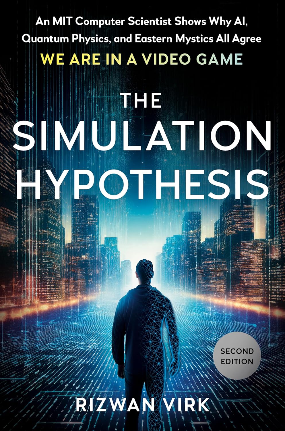
An MIT computer scientist connects AI, quantum physics and video games to take an uncomfortable question seriously: what if we live in a simulation? M
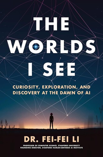
The memoir of the creator of ImageNet, the scientist who made the deep learning revolution in vision possible.
As an Amazon Associate, Zendoric earns from qualifying purchases; the price does not change for you. · all our recommended books →
🔗 Related on Zendoric
- Percival, Penn's AI that looks for hidden disease in every CT scan, still needs clinical validation · 2026-10-20
- An AI reads routine breast biopsies and predicts relapse almost as well as genomic tests costing thousands of dollars · 2026-07-10
- Meta patents an AI device that listens to, transcribes and analyzes your emotions to "personalize" workouts · 2026-07-10