When medical AI learns from the wrong organ: ovarian cancer study trained on thyroid images retracted

🕒 Published on Zendoric: October 8, 2026 · 00:57
✨ AI-generated · how it's made
On October 7, the journal Multimedia Tools and Applications retracted a 2024 study claiming to detect ovarian cancer with a neural network, after the ultrasound image bank turned out, according to the editors, to be of thyroids. The nine authors, spread across three countries and disciplines unrelated to gynecology, never responded. The case exposes how much clinical confidence can rest on data that was never verified.
📚 Go deeper

The only novel on the list, by a Nobel laureate: an artificial friend observes humans with a blend of lucidity and innocence no essay achieves.
As an Amazon Associate, Zendoric earns from qualifying purchases; the price does not change for you. · all our recommended books →
By Zendoric · October 8, 2026.
On October 7, 2026 the scientific journal Multimedia Tools and Applications retracted a study published in February 2024 that presented a convolutional neural network —the standard computer vision architecture, which learns to recognize visual patterns by filtering an image through successive layers— capable, according to its authors, of detecting and classifying ovarian cancer in ultrasound images. According to the retraction notice, the editors were unable to verify the origin of the dataset and reasonable doubts arose as to whether the images labelled as ovarian were in fact of the thyroid, a gland in the neck with a completely different anatomy on ultrasound.
The chain of responsibility, documented in the notice itself, is revealing. The dataset came, according to the original article, from a database described in a 2019 paper by Zhang and colleagues on the early detection of ovarian cancer using deep learning. That 2019 reference was itself retracted, on the same suspicion: that the images were of the thyroid. The 2024 study inherited the problem without anyone, along the way, checking that the images corresponded to the organ they claimed to depict.
The nine signing authors are affiliated with institutions in India, Saudi Arabia and the United States, in departments as disparate as data science, electrical engineering, botany, oral and maxillofacial surgery and biology, according to the article itself. The journal's Editor-in-Chief said he had lost confidence in the data and the conclusions, and noted that none of the authors responded to correspondence about the retraction. The notice does not allege specific misconduct beyond the impossibility of verifying the dataset, but that combination —affiliations with no apparent connection to gynaecological imaging and total silence towards the journal— is a pattern that research integrity watchdogs have long associated with paper mills, without that amounting here to a proven accusation.
The episode is not an isolated case. In recent years dozens of medical AI studies on chest X-rays, skin lesions and ultrasound have been retracted or corrected because of images that were mislabelled, duplicated between training and test sets, or outright unrelated to the disease they claimed to study. The underlying pattern is always the same: a neural network has no way of knowing that the label on its data is false. It will learn statistical regularities from whatever pixels it is handed and will produce impeccable confusion matrices and ROC curves, even if it is solving a different problem from the one announced.
Our reading is that this case confirms something we have already been pointing out in other areas of AI: a performance metric is a property of the data and of the process that produced it, not of the model. In the same way that a reasoning benchmark can be inflated depending on who runs the evaluation harness, near-perfect accuracy in cancer detection is worthless if nobody checked that the images were of the right organ. The bottleneck in medical AI lies not in architectural sophistication —already notably mature— but in the provenance, traceability and clinical verification of the data, an infrastructure far less eye-catching than training a model and one that receives a fraction of the funding and the prestige.
This does not invalidate the underlying promise. Ovarian cancer is diagnosed late precisely because current tools —the CA-125 test, transvaginal ultrasound— lack the sensitivity to detect it in time, and an AI system that reliably flagged suspicious scans for expert review would have real clinical value, consistent with the thesis that AI, in the long run, can bring us much earlier diagnoses and eliminate much of the harm that silent diseases cause today. But that horizon can only be built if the research community accepts the cost of verifying the provenance of every image with the same rigour with which it compares architectures, demands external validation at institutions other than those of origin, and treats authors' silence towards a journal as a warning sign, not a formality. The retraction of a single study does not hold back the field; the accumulation of studies built this way on never-verified data could, over time, erode the confidence that medical AI needs to deliver on its promise.
📚 Go deeper

An MIT computer scientist connects AI, quantum physics and video games to take an uncomfortable question seriously: what if we live in a simulation? M
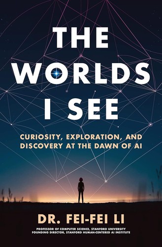
The memoir of the creator of ImageNet, the scientist who made the deep learning revolution in vision possible.
As an Amazon Associate, Zendoric earns from qualifying purchases; the price does not change for you. · all our recommended books →
🔗 Related on Zendoric
- What pigeons teach us about training AI to detect cancer · 2026-07-06
- Breast cancer leaves traces six years earlier: AI can already see them, and that forces a rethink of the entire screening process · 2026-06-28
- AI-generated CSAM at an Illinois school: the hardest abuse to detect no longer needs a hidden camera · 2026-07-04