Revived human retinas respond to light up to 10 hours after death, a step toward full eye transplants

🕒 Published on Zendoric: July 11, 2026 · 00:27
✨ AI-generated · how it's made
A team led by Eimear Byrne, of the Barcelona Institute of Science and Technology (BIST), has managed to keep donated human eyes metabolically active outside the body for up to 10 hours after death, double the time previously achieved by other scientists in 2022 (who had reached 5…
📚 Go deeper

The only novel on the list, by a Nobel laureate: an artificial friend observes humans with a blend of lucidity and innocence no essay achieves.
As an Amazon Associate, Zendoric earns from qualifying purchases; the price does not change for you. · all our recommended books →
A team led by Eimear Byrne, of the Barcelona Institute of Science and Technology (BIST), has managed to keep donated human eyes metabolically active outside the body for up to 10 hours after death, double the time previously achieved by other scientists in 2022 (who had reached 5 hours). The finding, published as a preprint on bioRxiv, is presented as a significant step toward the still-distant goal of whole-eye transplants capable of restoring vision.
The central problem this work addresses is the extreme fragility of the retina, the light-sensitive tissue located at the back of the eye. Unlike the cornea—whose transplantation is already an established practice that improves vision in people with corneal damage—the retina is connected to the central nervous system and is very sensitive to ischemia, that is, to the degeneration caused by a lack of oxygen. As Thomas Johnson, of Johns Hopkins University (who did not take part in the study), explains, even a brief period of ischemia probably causes irreversible degeneration of the light-sensitive neurons and circuits. This explains why, although a partial face and whole-eye transplant was already performed in 2023, it failed to restore the recipient's sight.
To reduce that damage, Byrne's team designed a system that inserts a flexible tube into the ophthalmic artery, which is responsible for supplying blood to the eye and surrounding structures. Through that tube they perfused the donated eye with an oxygenated solution, using a custom-developed device they have named the 'Eye-in-Care-Box,' equipped with sensors that automatically regulate pressure and flow, thereby mimicking the conditions the eye would experience inside the body.
Experimental validation was carried out in two phases. First, the researchers took both eyes from six donors, perfusing one of each pair and leaving the other unperfused as a control. The perfusion system preserved the retina's structure and maintained the health of the surrounding cells for up to 24 hours, while the unperfused eyes degraded quickly after their removal. In a second phase, they perfused another 36 donated eyeballs and found that 15 of their retinas produced electrical responses to light similar to those measured in living people, responses that were maintained for up to 10 hours after death. However, the article notes that it is unclear why the remaining 21 eyeballs did not show that response, suggesting that the technique is not yet consistent or fully understood.
Johnson stresses that this advance, although significant ('a tremendous achievement,' in his words), does not solve another major obstacle to restoring vision through a transplanted eye: the regeneration of the severed optic nerve fibers, needed for the eye to communicate with the brain's visual centers. Without that connection, he warns, a donated eye would have no way to transmit visual sensation to the recipient's brain. The article mentions that there are several groups researching how to promote optic-nerve regrowth, and Johnson suggests that the time has come to begin combining these promising interventions in the context of a whole-eye transplant.
Beyond its direct application to transplants, Byrne's own team points out that the Eye-in-Care-Box device could have value as a research tool in its own right: by keeping human eyes metabolically healthy outside the body, it would allow vision-related therapies to be tested directly on human tissue instead of resorting to animal models. Johnson agrees that there is potential to use this technology in developing new in vitro models and experimental paradigms for testing drugs and other therapies, as well as for better understanding ocular biology and pathology, with the advantage that the results would be more directly applicable to human disease and biology.
Overall, this is an incremental but concrete advance within a field—irreversible blindness from retinal diseases, such as age-related macular degeneration, which affects more than a million people in the United Kingdom—where current treatments only manage to slow the progression of the disease, without reversing it. The article itself is cautious: it does not claim that whole-eye transplantation is close to becoming a clinical reality, but rather that this perfusion technique reduces one of the technical obstacles (degeneration from ischemia) that until now made it unfeasible to preserve a donated eye in functional condition for long enough.
📚 Go deeper
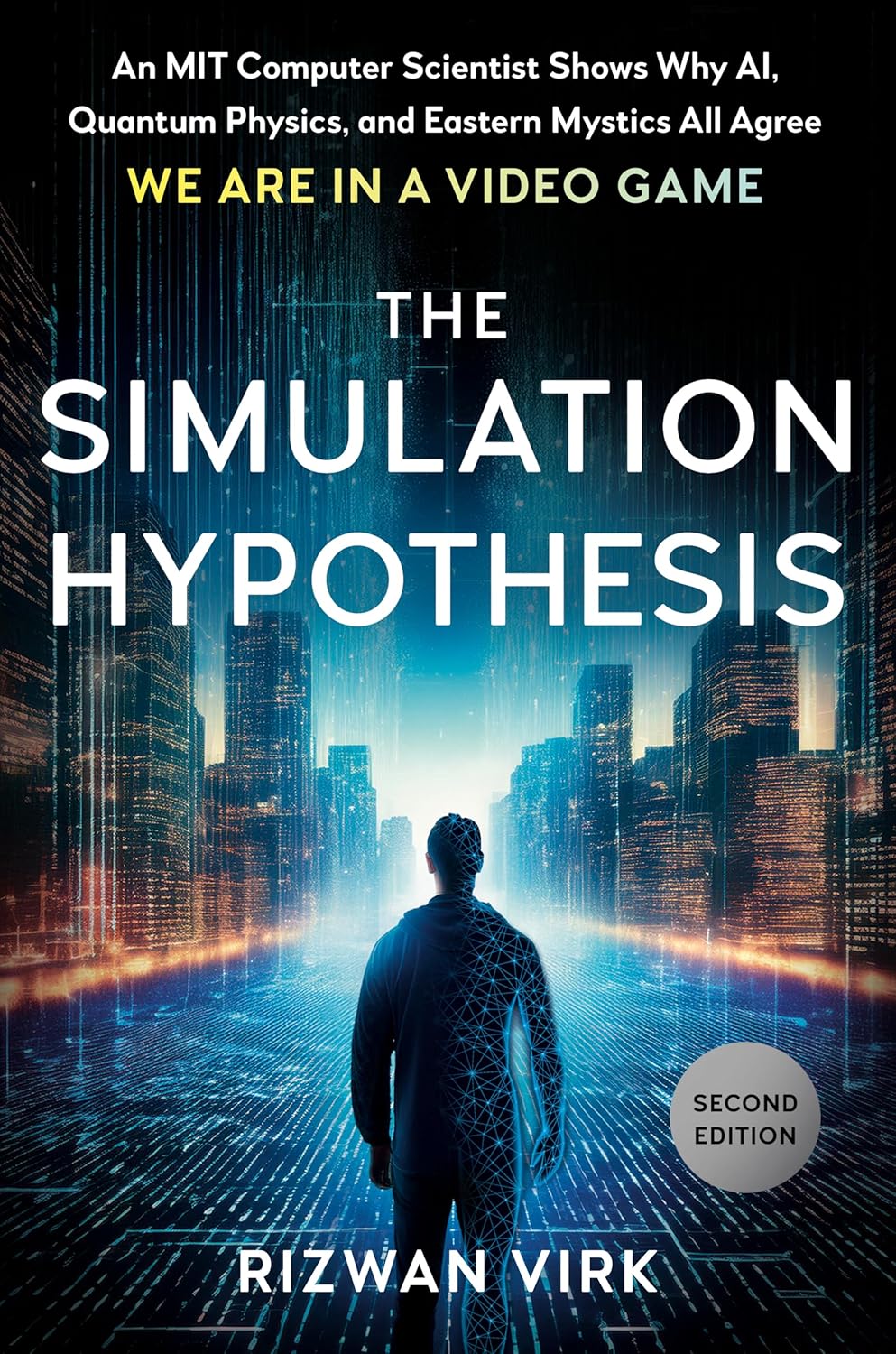
An MIT computer scientist connects AI, quantum physics and video games to take an uncomfortable question seriously: what if we live in a simulation? M
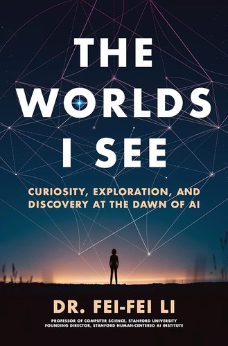
The memoir of the creator of ImageNet, the scientist who made the deep learning revolution in vision possible.
As an Amazon Associate, Zendoric earns from qualifying purchases; the price does not change for you. · all our recommended books →
🔗 Related on Zendoric
- AI against cancer: Harvard's model hits 94% accuracy and China tests a vaccine that hasn't yet reached human trials · 2026-07-21
- Cancer survivors in Washington launch an AI platform for oncology patients: the story, with few verifiable details · 2026-07-26
- The Pentagon formalizes more AI in combat decisions, and the market is already positioning itself · 2026-06-26